(+1) 302-319-8696
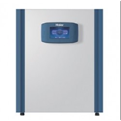
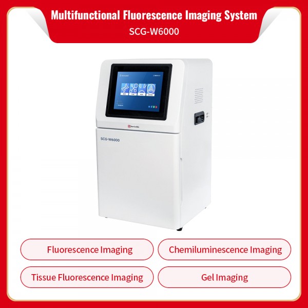
Servicebio Multifunctional Fluorescence Imaging System SCG-W6000
Equipped with four integrated imaging modules: fluorescence imaging, chemiluminescence imaging, tissue luminescence imaging, cell/microbial luminescence imaging and gel imaging. This all-in-one platform fully covers routine laboratory imaging applications.
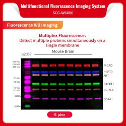
1. Fluorescent Western Blot Imaging Function: Fitted with three symmetric top-mounted fluorescence light sources at 470 nm, 530 nm and 650 nm.
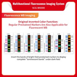
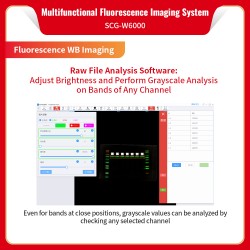
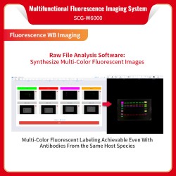
Enables simultaneous detection of multiple target proteins on a single membrane for multiplex fluorescent labeling; Proprietary image inversion function: standard pre-stained protein ladders can be directly applied for fluorescent WB assays. After inversion processing, bright ladder bands from brightfield are fully and clearly visible under darkfield; Dedicated raw data analysis software supports band brightness adjustment, grayscale quantification for each channel, and composite merging of multi-channel fluorescent images.
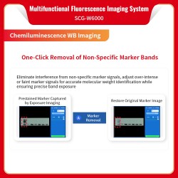
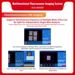
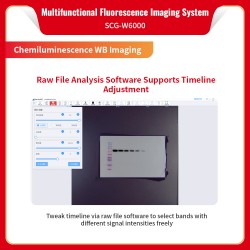
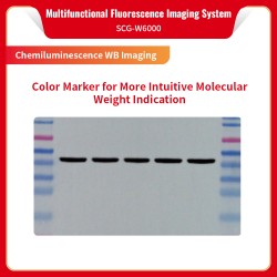
2. Chemiluminescent Western Blot Imaging Function:
Captures colored protein ladders with ultra-high detection sensitivity.
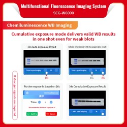
Accumulative exposure mode superimposes signal intensity on the base image to shorten operation time and rapidly yield optimal WB results.
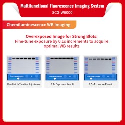
Fine exposure adjustment in 0.1-second increments for precise capture of crisp, well-resolved bands.
One-click ladder removal to restore authentic target band signals.
Supports simultaneous exposure of multiple membranes; raw files can be split into independent single-membrane files for separate analysis to boost experimental throughput.
The raw data analysis software features an adjustable timeline, allowing users to freely select band signals of varying intensities according to experimental requirements.
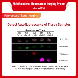
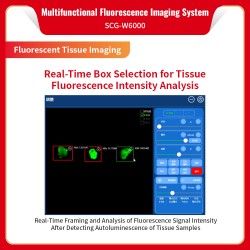
3. Tissue Fluorescence Imaging:
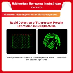
Captures autofluorescence signals of tissue specimens; real-time frame selection enables quantitative analysis of tissue fluorescence intensity. Signal presence and magnitude guide subsequent experiments efficiently and improve workflow productivity; Rapid assessment of fluorescent protein expression in cell culture plates and bacterial agar plates, meeting research demands for cell and microbial studies.
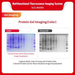
4.Gel Imaging Function:
Protein Gel Imaging: Supports both color and grayscale protein gel acquisition. Color gel outputs deliver more intuitive visual results.
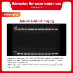
Nucleic Acid Gel Imaging: Comes standard with 254 nm, 310 nm and 365 nm LED UV light sources, compatible with samples labeled with common nucleic acid dyes including EB, SerRed, SerBlue, GoldView and SYBR Green. LED UV illumination delivers uniform lighting, superior image quality and extended service life.
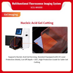
Nucleic Acid Gel Band Excision: Supplied with a standard UV laser safety shield with optical density cutoff > OD7, offering high-grade UV protection for secure gel cutting operations.
Parameters
| Product Name | Multifunctional Fluorescence Imaging System | |
| Cat.No. | SCG-W6000 | |
| Dimensions | 400mm*371mm*700mm | |
| Camera | Pixel | 900万 |
| Resolution | 3000x3000 | |
| Pixel Size | 3.76umx3.76um | |
| Target Size | 1“(11.28mmx11.28mm) | |
| Full Well Capacity | 16.5ke-(HCG),50.5ke-(LCG) | |
| Sensitivity | 877mv@1/30s | |
| Readout Noise | 1.24e-(HCG),3.22e-(LCG) | |
| Dark Current | 0.0003e-/s/pixel@-15℃ | |
| Signal-to-Noise Ratio | 42.2dB(HCG), 47dB(LCG) | |
| Exposure Time | 0.1ms~1h | |
| Binning Mode | 1x1,2x2,3x3 | |
| Grayscale | 16-bit (65536 gray levels) | |
| Cooling | -40°C relative to ambient | |
| Type | Monochrome Camera | |
| Lens | Aperture | F0.95-F16 |
| Focal Length | 17mm | |
| Type | Motorized Lens | |
| Light Sources | Brightfield | Episcopic LED white light, symmetrical on both sides |
| UV | 310 nm LED array, uniform transmission illumination | |
| Episcopic 254 nm/365 nm LED UV light, symmetrical on both sides | ||
| Fluorescence | Episcopic fluorescence light sources: 470 nm, 530 nm, 650 nm, symmetrical on top | |
| Blue/White Dual Light | Optional: blue/white transmission switch, 3 power levels | |
| Dark Box | Light-tightness | Fully light-tight, isolates ambient light |
| Door sensor | Controls brightfield and 310 nm UV LED sources | |
| Filter wheel | Switches filters according to mode to match chemiluminescence, gel, and fluorescence applications | |
| Field of View | Blotting membrane effective field of view: 155 mm x 155 mm | |
| Protein gel effective field of view: 155 mm x 155 mm | ||
| Nucleic acid gel effective field of view: 215mm*215mm | ||
| Fluorescent membrane effective field of view:136mm*136mm | ||
| Gel Cutting | When door is opened, UV light source can be pulled out; use UV protective plate for gel cutting | |
| Software Functions | Auto Exposure | Smart exposure quickly determines optimal exposure time, auto binning, combined with timeline imaging and time accumulation; one operation yields optimal image |
| Manual Exposure Mode | High Quality: highest image quality | |
| Standard: balances image quality and speed | ||
| High Sensitivity: fastest exposure speed | ||
| Real-Time Imaging | Shows signal changes during exposure, captures every detail; overexposed areas are highlighted | |
| Temporal Imaging | Generates every frame during exposure; user can select any frame as final output via precise backtracking | |
| Time Accumulation | For underexposed samples, user can continue exposure after the initial exposure ends, adding extra exposure time | |
| Marker Removal | When markers show extra signal bands, one-click removal without re-preparing the sample | |
| Industrial Computer | 10.4-inch, 1024x768, Windows 10, 16 GB RAM, 512 GB SSD, built-in Bluetooth and Wi-Fi | |
| External Interfaces | USB3.0 x2 | |
| Operating Voltage | 90-132 VAC / 180-264 VAC (selectable via switch), 47-63 Hz | |
| Power | ≤300W | |
| Net Weight | 35 Kg | |
Fully functional, integrating four core modules: fluorescence imaging, chemiluminescence imaging, tissue fluorescence imaging and gel imaging. It meets diverse experimental requirements, including fluorescent WB imaging, chemiluminescent WB imaging, tissue fluorescence observation, detection of fluorescent protein expression in cells and microorganisms, as well as imaging and band excision of protein gels and nucleic acid gels.