(+1) 302-319-8696
Product Information
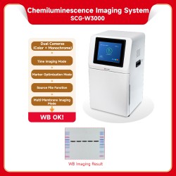
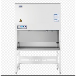
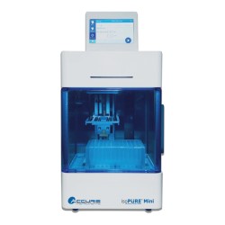
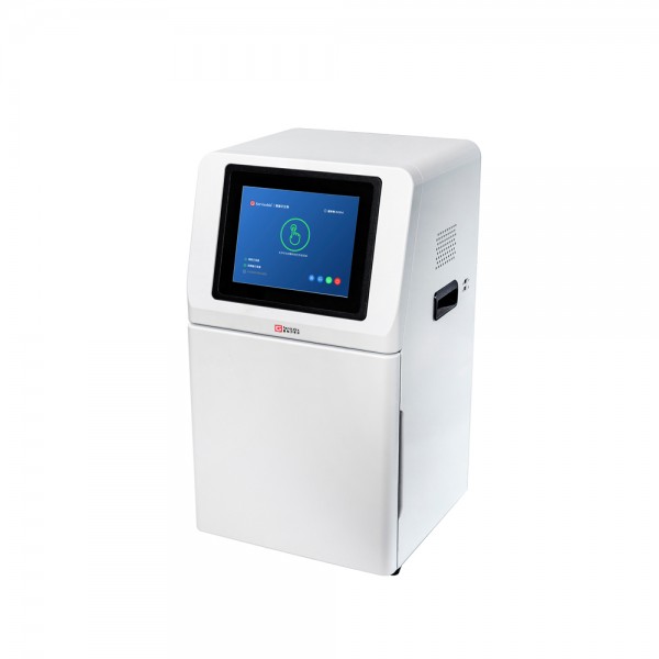
SCG-W3000 PLUS Chemiluminescence Imaging System
A digital imaging system capable of preserving the complete signal timeline.
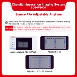
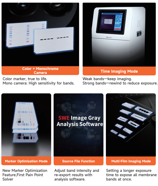
Using the "Source File Analysis Software", you can adjust the timeline and selectively capture signals from bands of varying intensities.
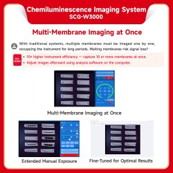
Temporal Imaging:
1. Even weak membranes can produce WB results in a single run using cumulative exposure mode.
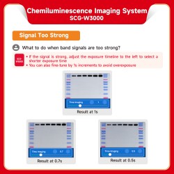
2. For overexposed strong membranes: fine-tune in 0.1 s increments to achieve optimal WB results.
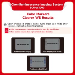
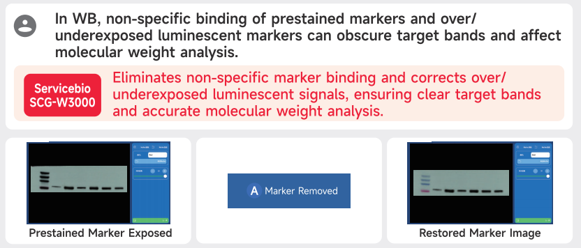
Marker Removal:
3. For non-specific binding signals from the marker: one-click restoration to reveal true marker bands.
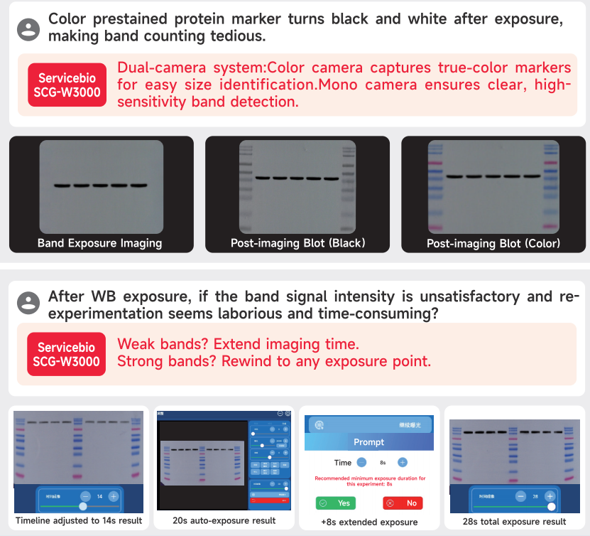
4. Dual cameras: a color camera for marker imaging, providing true-to-life colors; a monochrome camera for band imaging, offering high sensitivity and excellent performance for weak signals.
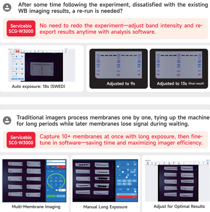
5. Supports simultaneous imaging of 10 or more membranes, greatly improving instrument efficiency – one machine, 10x performance.
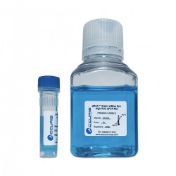
Paired with the ECL Chemiluminescent Substrate Reagent Combo Pack(G2161-200ML), it meets exposure requirements for high, medium, and low concentration samples. Experience a better WB imaging process and obtain superior WB imaging results!
SWE Imaging Grayscale Analysis Software-SCG-W2000 SCG-W3000-3.3.2-win32-x64-setup.exe
Download link from Google Drive:
https://drive.google.com/file/d/1IoXazML9PDHoN4HqOYm-t3C2bVFvQ_rt/view?usp=drive_link
SOFTWARE USER MANUAL:
SWE Imaging Grayscale Analysis Software-SCG-W2000 SCG-W3000.pdf
| Product Name | Chemiluminescence Imaging System | |
|---|---|---|
| Cat. No. | SCG-W3000 PLUS | |
| Dimensions | 400mm×371mm×700mm | |
| Camera 1 | Pixel Resolution | 9 million pixels |
| Resolution | 2992*3000 | |
| Pixel Size | 3.76μm×3.76μm | |
| Target Size | 1 “(11.28mm×11.28mm) | |
| Full Well Capacity | 16.5ke-(HCG),50.5ke-(LCG) | |
| Sensitivity | 877mv@1/30s | |
| Readout Noise | 1.24e-(HCG), 3.22e-(LCG) | |
| Dark Current | 0.0003e-/s/pixel@-15℃ | |
| Signal-to-Noise Ratio | 42.2dB(HCG), 47dB(LCG) | |
| Exposure Time | 0.1ms~1h | |
| Binning Mode | 1×1, 2×2, 3×3 | |
| Grayscale | 16-bit (65536 levels) | |
| Cooling | Relative to Ambient Temperature-40℃ | |
| Camera Type | Black and White Camera | |
| Camera 2 | Pixel Resolution | 45 million pixels |
| Resolution | 2992*3000 | |
| Pixel Size | 2.315×2.315μm | |
| Target Size | 1.4“(18.93×13 mm) | |
| Full Well Capacity | 10.8ke- | |
| Sensitivity | 419mv | |
| Readout Noise | 2.12e- | |
| Dark Current | 0.12mV | |
| Signal-to-Noise Ratio | 40.3dB | |
| Exposure Time | 17μs~15s | |
| Binning Mode | 1×1, 2×2, 3×3 | |
| Grayscale | 8bit(256 gray) | |
| Camera Type | Color Camera | |
| Lens1 | Aperture | F0.95-F16 |
| Focal Length | 17mm | |
| Type | Prime lens | |
| Light Source | Bright Field Light Source | Downward-facing LED white light source, symmetrically distributed on both sides |
| Dark Box | Light Isolation | Fully light-sealed, isolates environmental light. |
| Door Control | Door control sensor can control the on/off of the bright field light source. | |
| Field of View | Effective field of view is 136mm*136mm (expandable to 200mm×200mm if needed). | |
| Auto Exposure | Intelligent exposure technology can quickly determine the optima | |
| exposure time and automatically perform binning. Combined with | ||
| time-lapse imaging and time accumulation functions, users can | ||
| achievethe bestimage results withjust one opematchration. | ||
| Software Functions | Exposure Modes | High Quality:Highest image quality |
| Standard:Balancesimage qualityand exposurespeed | ||
| High Sensitivity:Fastestexposure speed | ||
| Real-time lmaging | Real-time presentation of the changes in sample signals during the | |
| exposure process, allowing for the observation of every detail ofthe | ||
| capture. Overexposed areas will be indicated for samples with | ||
| overexposure. | ||
| Time Imaging | After exposure is complete, each frame image within the exposure | |
| time can be generated.Through precise retrospective adjustments | ||
| users can choose any frame image within that exposure time as the | ||
| final output. | ||
| Time Accumulation | For samples with insufficient exposure, users can choose to continue | |
| exposure after the initial exposureis completed, enabling the sample | ||
| to receive additionalexposure on topofthe already exposed time. | ||
| Optional Settings | Color Marker/Black & White Marker, Overexposure Prompt/No | |
| OverexposurePrompt | ||
| Industrial Computer | 10.4-inch display with a resolution of 1024x768, running on Windows 10 operating | |
| system,featuring16GBofRAM,512GBssD,and built-in Bluetooth and WiFi. | ||
| External Interfaces | 2 USB 3.0 ports | |
| Operating Voltage | 90~132VAC/180~264VAC(selectable via switch),47~63Hz. | |
| Product Power | ≤200W | |
| Product Net Weight | 23.45Kg | |
Notes:
Do not touch or scratch the internal lens of the dark box with hands or sharp objects.
After placing the sample, ensure the instrument door is properly closed to prevent external light from entering and affecting results.
During imaging, avoid shaking the workbench or instrument to prevent compromised image quality.
Observe electrical safety—do not pull the power cord or move the instrument during operation.
After experiments, clean the dark box thoroughly, removing all samples and residues.
| Cat. No. | SCG-W5000 PLUS | SCG-W3000 PLUS | SCG-W1000 PLUS |
|---|---|---|---|
| Dimension | 400×371×700 mm | 400×371×700 mm | 400×371×700 mm |
| Camera | Depth-cooled high sensitivity camera | Depth-cooled high sensitivity camera | High-sensitivity camera |
| Resolution | 2992×3000,9 megapixels | 2992×3000,9 megapixels +2992×3000 | 3072×2048,6.3 megapixels |
| 45 megapixels | |||
| Pixel | 3.76×3.76 μm | 3.76×3.76 μm | 2.4×2.4 μm |
| Shooting Area | Effective field of view for blotting film/protein gel: 136×136 mm | Blotting Film | Nucleic Acid Gel / Protein Gel |
| Effective field of view for nucleic acid gel: 140×140 mm. | 136×136 mm | 140×140 mm | |
| Cooling Temperature | Relative ambient temperature -40°C | Relative ambient temperature -40°C | - |
| Light Source | Bright-field Light Source: Downward-facing LED white light source, symmetrically distributed on both sides. | Downward-facing LED white light, symmetrically distributed on both sides | Bright-field Light Source: Downward-facing LED white light source, symmetrically distributed on both sides. |
| UV Light Source: 310 nm LED array for uniform transmission illumination. | UV Light Source: 310 nm LED array for uniform transmission illumination. | ||
| Industrial Computer | 1match0.4 inches, 1024×768 | 10.4 inches, 1024×768 | 10.4 inches, 1024×768 |
| Windows operating system | Windows operating system | Windows operating system | |
| External Interface | 2 USB3.0 | 2 USB3.0 | 2 USB3.0 |
| Working Voltage | 90~132VAC/180~264VAC | 90-132V/180-264V | 90~132VAC/180~264VAC |
| (Selectable via switch)47~63HZ | (Selectable via switch)47~63HZ | ||
| Product Power | 200 W | ≤200 W | 200 W |
| Net Weight | 25 kg | 23.45 kg | 30 kg |
| Real-Time Imaging | Yes | Yes | - |
| Time Imaging | Yes | Yes | - |
| Time Accumulation | Yes | Yes | - |
| Auto Exposure | Yes | Yes | Yes |
| Choice of 3 Imaging Modes | Yes | Yes | - |
| Protein Gel/Nucleic Acid Gel Imaging | Yes | - | Yes |
| Nucleic Acid Gel Cutting | Yes | - | Yes |