(+1) 302-319-8696
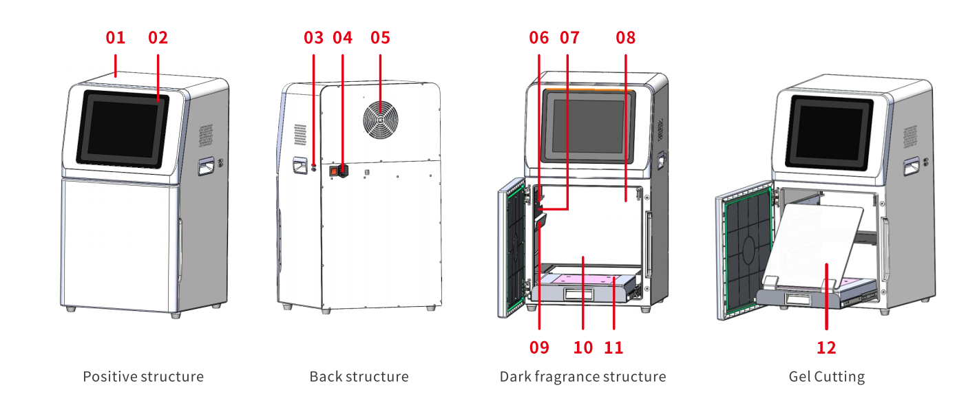
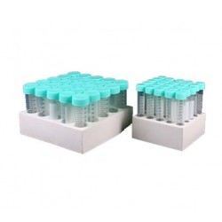
Product Information
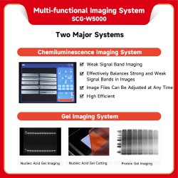
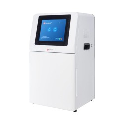
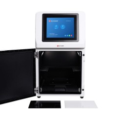
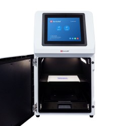
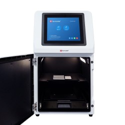
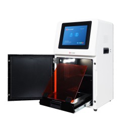
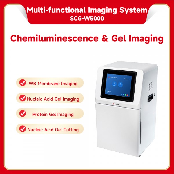
The SCG-W5000 PLUS Multifunctional Imaging System — capable of chemiluminescence imaging, gel imaging, and gel cutting.
1.One-shot imaging for WB results regardless of signal strength
Clear Western blot results can be obtained in a single acquisition, whether the signal is strong or weak.
For weak signals: exposure time can be extended directly from the original acquisition to continue imaging.
For strong signals: any image can be retrieved from the time course within the recorded exposure sequence.
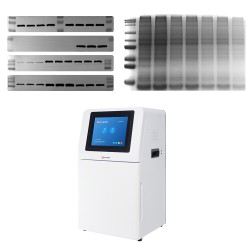
2. Supports simultaneous imaging of up to 10 membranes—or even more—greatly improving instrument efficiency, with one system, 10x performance.
3. Capable of imaging color Marker with high sensitivity, delivering excellent performance even for weak signals.
4.Marker optimization function, eliminates non-specific bands caused by interactions between pre-stained markers and antibodies, and allows adjustment of overexposed or underexposed luminescent marker signals.
5.Adjustable raw data files, raw image files are saved for flexible post-acquisition adjustment. Band thickness can be optimized at any time on a computer, ensuring ideal results even after the experiment.
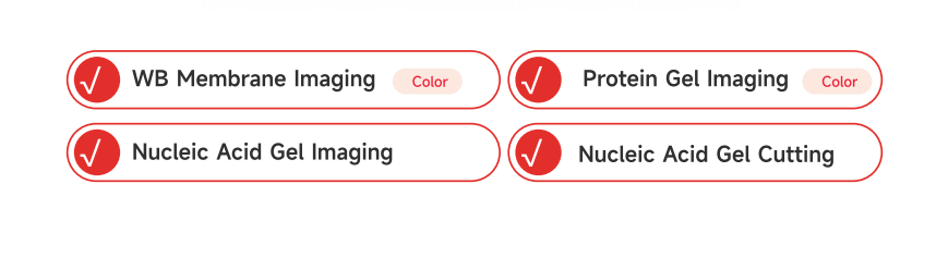
6.Protein gel imaging: Supports both color and grayscale imaging of protein gels. Color images provide more intuitive and informative results.
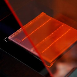
7.Nucleic acid gel imaging: Equipped with standard 310/254/365 nm LED UV light sources, compatible with common nucleic acid stains such as EB, SerRed, SerBlue, GoldView, and SYBR Green.
LED UV illumination ensures more uniform lighting, improved image quality, and longer service life.
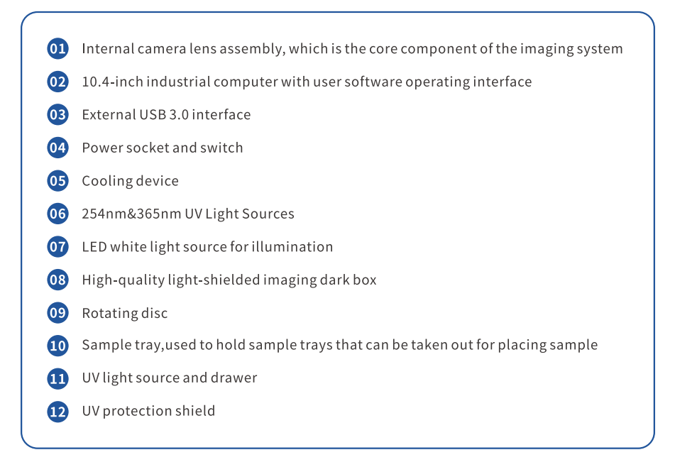
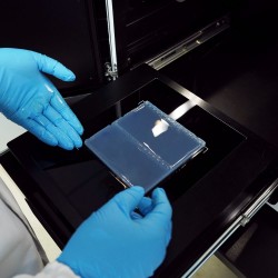
8.Safe nucleic acid gel excision: Comes standard with a UV laser protection shield (cutoff depth > OD7), providing a high level of safety for gel cutting operations.
Compatible with ECL Chemiluminescent Substrate Reagent Combo Pack (G2161-200ML), enabling optimal exposure for samples across high, medium, and low concentrations. Delivers an enhanced Western blot imaging experience and superior results.
Technical Specifications
| Product Name | Multifunctional Imaging System | |
| Cat.No. | SCG-W5000 PLUS | |
| Dimensions | 400×371×700 mm | |
| Camera | Pixel Resolution | 9 Megapixels |
| Resolution | 2992×3000 | |
| Pixel Size | 3.76×3.76μm | |
| Target Size | 1“(11.28×11.28 mm) | |
| Full Well Capacity | 16.5ke-(HCG),50.5ke-(LCG) | |
| Sensitivity | 877mv@1/30s | |
| Readout Noise | 1.24e-(HCG),3.22e-(LCG) | |
| Dark Current | 0.0003e-/s/pixel@-15℃ | |
| Signal-to-Noise Ratio | 42.2dB(HCG),47dB(LCG) | |
| Exposure Time | 0.1ms~1h | |
| Binning Mode | 1×1,2×2,3×3 | |
| Grayscale | 16-bit(65536 levels) | |
| Cooling | Relative to Ambient Temperature -40°c | |
| Camera Type | Black and White Camera | |
| Lens | Aperture | F0.95-F16 |
| Focal Length | 17mm | |
| Type | Motorized zoom lens | |
|
Light Source |
Bright Field Light Source |
Downward-facing LED white light source, symmetrically distributed on both sides |
| Ultraviolet Light Source | 310nm LED array with uniform transmitted illumination,254nm/365nm LED ultraviolet light sources (symmetrically distributed on both sides) |
|
| Dark Box | Light Isolation | Fully light-sealed, isolates environmental light. |
| Door Control |
The door control sensor can control the on/off of the bright field light source. |
|
| Rotating Disc |
Switch the filter according to the current mode to match the applications of chemiluminescence and gel imaging. |
|
| Field of View |
Effective field of view for membrane imaging is 140mmx 140mm Effective field of view for protein gel imaging is 140mmx 140mm The effective field of view for nucleic acid gel imaging is 140mmx140mm |
|
| Gel Cutting |
After opening the door, the UV light source can be extracted and used with a UV protective board for cutting adhesive |
|
|
Software Functions |
Exposure Modes |
High Quality: Image quality is the highest |
| Auto Exposure |
Intelligent exposure technology quickly determines the optimal exposure time. With the combination of time imaging and time accumulation functions, users can achieve the best image results with just one operation. |
|
| Real-time Imaging |
Real-time presentation of the changes in sample signals during the exposure process, allowing for the observation of every detail of the capture. Overexposed areas will be indicated for samples with overexposure. |
|
| Time Imaging |
After exposure is complete, each frame image within the exposure time can be generated Through precise retrospective adjustments, users can choose any frame image within that exposure time as the final output. |
|
| Time Accumulation |
For samples with insufficient exposure, users can choose to continue exposure after the initial exposure is completed, enabling the sample to receive additional exposure on top of the already exposed time. |
|
| Industrial Computer | 10.4"display(1024x768)Windows 10 0s 16GB RAM,512GB SSD,Integrated Bluetooth/Wi-Fi |
|
| External Interfaces | USB 3.0×2 | |
| Operating Voltage | 90~132VAC/180~264VAC(selectable via switch),47~63Hz | |
| Product Power | ≤300W | |
| Product Net Weight | 30.65Kg | |
Notes
It is prohibited to touch or scratch the internal lenses of the dark box with hands or sharp objects;
After placing the experimental samples, make sure to close the instrument's flip door to prevent external light from entering
the dark box and affecting the experimental results;
During imaging experiments, shaking the experimental table or instrument is prohibited to avoid impacting the image
quality;
Pay attention to electrical safety. Pullingor moving the power cord during the experiment is prohibited;
After the experiment is completed, clean the samples and any residues inside the dark box thoroughly.
| Cat.No. | SCG-W3000 PLUS | SCG-W5000 PLUS | SCG-W2000 PLUS | SCG-W1000 PLUS |
| Dimension | 400×371×700 mm | 400×371×700 mm | 400×371×700 mm | 400×371×700 mm |
| Camera |
Depth-cooled high sensitivity color camera | Depth-cooled high sensitivity camera |
Depth-cooled high sensitivity camera |
Color Camera |
| Resolution | 2992×3000,9 megapixels |
2992×3000,9 megapixels |
2992×3000,9 megapixels |
3072×2048,6.3 megapixels |
| Pixel | 3.76×3.76 μm | 3.76×3.76 μm | 3.76×3.76 μm | 2.4×2.4 μm |
| Shooting Area |
Blotting Membrane Effective field of view: 136×136 mm |
Blotting Membrane/Protein Gel/Nucleic Acid Gel Effective field of view: 140×140 mm. |
Blotting Membrane: 136×136 mm (Expandable up to 200×200 mm if needed) |
Nucleic Acid Gel /Protein Gel Maximum Effective field of view:140×140 mm |
Cooling Temperature |
Relative ambient temperature -40℃ |
Relative ambient temperature -40℃ |
Relative ambient temperature -40℃ |
一 |
| Light Source | Downward-facing LED white light, symmetrically distributed on both sides |
Brightfield Light Source:Downward- facing LED white light source,symmetrically distributed on both sides. UV Light Source:310nm LED array for uniform transmission illumination. Downward-facing 254 nm /365nm LED UV Light Source,symmetrically distributed on both sides. |
Brightfield Light Source:Downward-facing LED white light source, symmetrically distributed on both sides. |
Brightfield Light Source:Downward-facing LED white light source,symmetrically distributed on bot sides. UV Light Source:310 nm LED array for uniform transmission illumination. Downward-facing 254 nm/365nm LED UV Light Source,symmetrically distributed on both sides. |
| Industrial Computer |
10.4 inches,1024×768 Windows 10 operating system | 10.4 inches,1024×768 Windows 10 operating system | 10.4 inches,1024×768 Windows 10 operating system | 10.4 inches,1024×768 Windows 10 operating system |
| External Interface |
2 USB3.0 | 2 USB3.0 | 2 USB3.0 | 2 USB3.0 |
| Working Voltage | 90-132V/180-264V | 90~132VAC/180~264VAC(Selectable via switch)47~63HZ | 90~132VAC/180~264VAC(Selectable via switch)47~63HZ | 90~132VAC/180~264VAC(Selectable via switch)47~63HZ |
| Product Power | ≤200 W | ≤300 W | 100 W | ≤300 W |
| Net Weight | 23.45 kg | 30.65 kg | 22 kg | 30.65 kg |
| Real-Time Imaging |
Yes | Yes | Yes | 一 |
| Time Imaging | Yes | Yes | Yes | 一 |
| Time Accumulation |
Yes | Yes | Yes | 一 |
| Auto Exposure | Yes | Yes | Yes | Yes |
| Choice of 3 Imaging Modes |
Yes | Yes | Yes | 一 |
| Protein Gel/ Nucleic Acid Gel Imaging |
一 | Yes | 一 | Yes |
| Nucleic Acid Gel Cutting |
一 | Yes | 一 | Yes |