 Hotline Free:
(+1) 302-319-8696
| Specification | Details |
|---|---|
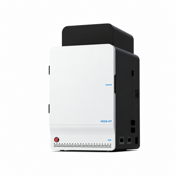
| Model | MOIS HT |
| Product Type | Small Animal In Vivo Optical Imaging System |
| Imaging Modules | Bioluminescence Imaging, Fluorescence Imaging, Spectral High-Resolution Imaging |
| Detector Type | Back-Illuminated Scientific-Grade CCD |
| CCD Cooling Temperature | -90°C |
| CCD Resolution | 1024 × 1024 Pixels |
| Quantum Efficiency | ≥90% (500–700 nm) |
| Readout Noise (Typical) | 2.9 e⁻ at 50 kHz; 6.6 e⁻ at 1 MHz |
| Lens Aperture | F-Stop ≤0.95 |
| Minimum Field of View | 2.5 × 2.5 cm (Optional) |
| Maximum Field of View | 25 × 25 cm |
| Spectral Coverage | 400–900 nm |
| Excitation Filters | 19 Narrow-Band Filters |
| Emission Filters | 7 Narrow-Band Filters |
| Broadband Light Source | 150 W Halogen-Tungsten |
| Positioning System | Guide Laser with Real-Time FOV Center Display |
| Multi-Animal Imaging Chamber | 5 Channels |
| Animal Imaging Capacity | At Least 5 Small Animals Simultaneously |
| Chamber Temperature Control | 20–40°C; Accuracy ±0.1°C |
| Anesthesia Gas Control | Isoflurane, Adjustable Concentration 0.5–5% |
| Anesthesia Accessories | 5-Channel Anesthesia Tray and Active Gas-Scavenging Masks |
| Real-Time Acquisition | Real-Time Imaging, Time-Series Acquisition, Multimode Switching, Synchronous Acquisition Support |
| Image Analysis | Image and Time-Spectral Analysis; Advanced Molecular Quantification Requires Software Authorization |
| Calibration | NIST-Traceable Light Calibration and Excitation Uniformity Correction |
| Upconversion Fluorescence Imaging (UCFI) | Expandable / Upgradeable |
| X-Ray Imaging Module | Not Included |
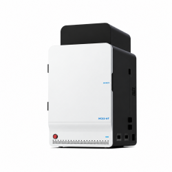
The CravitySci MOIS HT Small Animal In Vivo Imaging System is a highly sensitive, multimodal optical imaging platform designed for non-invasive visualization, monitoring, and quantitative analysis of biological processes in preclinical research. Integrating bioluminescence, fluorescence, and spectral high-resolution imaging technologies, the system enables researchers to investigate molecular and cellular activity in living small animals with minimal experimental disruption.
Designed for demanding low-light applications, the MOIS HT combines deep-cooled CCD detection, high-efficiency optics, advanced spectral separation, and comprehensive image optimization. Its flexible imaging capabilities support research ranging from localized signal detection to whole-body monitoring, including tumor development, immune and stem cell tracking, gene expression, drug distribution, and therapeutic response evaluation.
The integrated imaging chamber, environmental control system, and data acquisition software provide a versatile platform for multi-animal studies and repeated observations across different experimental conditions and time points.
The MOIS HT is optimized for detecting extremely weak optical signals in living biological subjects. Its deeply cooled back-illuminated CCD detector minimizes thermally generated background noise, while the high-efficiency optical system maximizes photon collection. These technologies work together to enhance signal-to-noise performance and produce detailed images for demanding bioluminescence, fluorescence, and molecular imaging applications.
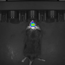
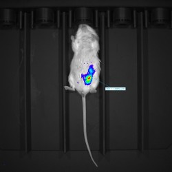
Bioluminescence imaging enables non-invasive detection of light emitted from biological reporters in living subjects. The highly sensitive optical detection system supports monitoring of tumor growth, gene expression, cellular activity, and treatment responses, including experiments requiring detection of low-intensity signals.
Fluorescence imaging enables visualization of labeled cells, tissues, and molecular targets using compatible fluorescent probes. The optical system accommodates commonly used fluorescence wavelengths, supporting applications such as cell migration, molecular targeting, biodistribution, and disease-associated biomarker studies.
Advanced spectral unmixing technology helps resolve signal overlap between different fluorescent probes while suppressing interfering background fluorescence. This capability enables more accurate identification and localization of individual fluorescence signals within complex biological environments, supporting multi-color imaging and molecular co-localization studies.
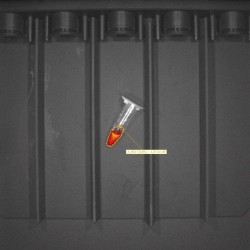
The MOIS imaging platform is described in the product literature as supporting Cerenkov Luminescence Imaging (CLI) for radionuclide-based optical studies. CLI uses light generated through interactions associated with suitable radionuclide tracers, enabling investigation of radiotracer distribution and selected drug evaluation applications. The exact CLI module and software configuration should be confirmed for the supplied system.
The MOIS HT incorporates optical calibration and image-processing technologies designed to improve measurement consistency and reduce unwanted background interference. Its specialized light-tight imaging chamber helps minimize external light contamination during sensitive signal acquisition.
The integrated acquisition software supports real-time image capture, time-series experiments, communication connectivity, and convenient switching between optical imaging modes. Synchronous acquisition functionality is also supported, enabling flexible experimental data collection.
Image analysis and time-spectral analysis tools assist researchers in interpreting experimental signals and comparing imaging results. Additional molecular quantitative analysis functions may require separate software authorization.
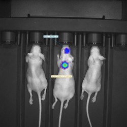
Designed for efficient preclinical workflows, the system accommodates simultaneous imaging of multiple small animals. An integrated temperature-control environment supports controlled imaging conditions, while the multi-channel anesthesia arrangement allows inhalation anesthesia during imaging procedures.
Active anesthetic gas-scavenging masks help limit waste anesthetic gas leakage, and crosshair positioning lasers assist with accurate placement of experimental subjects. These features support consistent positioning, efficient handling, and repeated observation in longitudinal studies.
The manufacturer provides examples of imaging experiments and data analysis applications, including:
The MOIS HT configuration supports bioluminescence, fluorescence, and spectral high-resolution imaging. Upconversion fluorescence imaging is identified as an expandable or upgradeable capability rather than a confirmed standard module. Certain advanced analysis functions require software authorization.
X-Ray imaging is not included in the MOIS HT configuration. Availability of specialized imaging modules, associated software, additional accessories, and the final supplied configuration should be confirmed before ordering.